Rings, Dots, Lines & Spots in Ophthalmology: The Ultimate Clinical Guide
Description
Course Description
Welcome to Rings, Dots, Lines & Spots in Ophthalmology | Dr. Jay Daxini.
If your goal is to master essential ophthalmic clinical signs with Dr. Jay Daxini's visual guide, you have found the perfect resource. Explore key rings, spots, and lines, and prep with the Ophtha MCQ app!
In Rings, Dots, Lines & Spots in Ophthalmology: The Ultimate Clinical Guide, we provide a high-yield, visually driven breakdown of the most crucial clinical findings. Accurately identifying these subtle signs is a core competency for any eye surgeon. This course covers everything from corneal iron deposits to retinal manifestations, ensuring you recognize every critical sign in clinical practice.
What You Will Master
RINGS IN OPHTHALMOLOGY
Ring Keratitis: Acanthamoeba is a rare parasitic infection of the cornea that primarily occurs in contact lens wearers. You will learn to identify the corneal ring infiltrate seen in patients with Acanthamoeba keratitis.
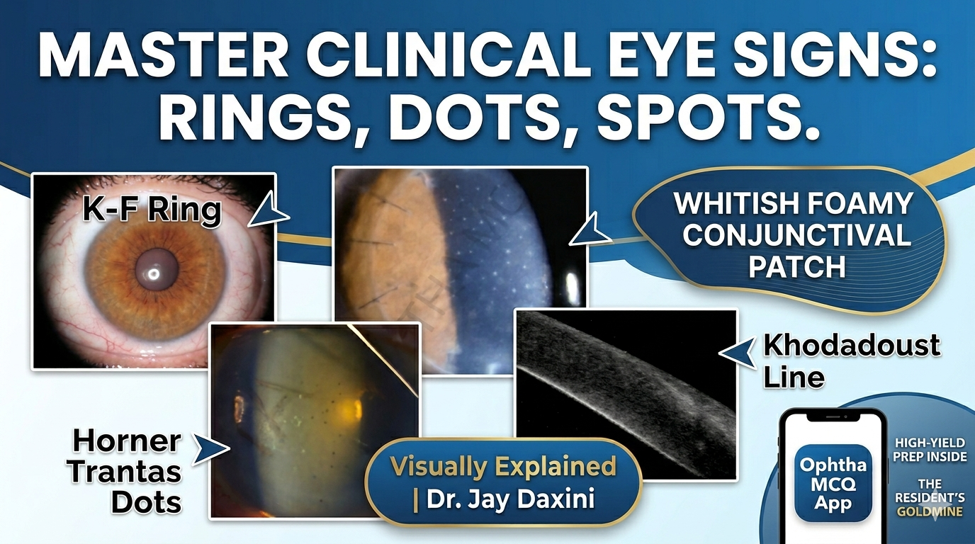
Kayser-Fleischer's Ring: Classic corneal sign.
Corneal Rust Ring: A small, reddish brown, circular opacity remained in the cornea after the removal of an iron foreign body.
Coat's White Ring: A form of iron deposit at the level of Bowman's layer commonly associated with previous corneal foreign body.
Fleischer's Ring: Fleischer rings are corneal rings. Iron deposits in your cornea cause Fleischer rings. This type of ring is related to keratoconus, a condition that causes your eye to bulge out in a cone shape instead of the usual round shape of your cornea. It is visible all around the base of cone in keratoconus.
Soemmering's Ring: Early opacification of lens capsule after cataract extraction.
Vossius Ring: Iris pigment on anterior lens capsule in concussion injury to eye.
Weiss Ring: A Weiss ring is a vitreous opacity, or floater, that has a distinctive ring, oval, or C-shape. It's a piece of glial tissue that remains attached to the optic nerve head after the vitreous gel separates from it. Its appearance is a sign that a complete posterior vitreous detachment has occurred.
Double Ring Sign: The peripheral margin of the encircling ring corresponding to the border of a normal-sized optic disc. Seen in hypoplasia of the optic disc.
Golden Ring Sign: Golden ring within the lens is evidence of successful delineation.
SPOTS IN OPHTHALMOLOGY
Krachmer Spots: Subepithelial infiltrates seen in corneal stromal graft rejection.
Elschnig's Spots: Black spots surrounded by bright yellow or red halos in patients with advanced hypertensive retinopathy.
Bitot's Spot: A Bitot's spot is a foamy, whitish, triangular patch on the white of the eye (conjunctiva), classically a sign of Vitamin A deficiency (VAD), indicating xerophthalmia. Vitamin A deficiency can also appear with other issues like malabsorption, liver disease or even normal Vitamin A levels due to genetic or environmental factors. It is a white, foamy area of keratinising squamous metaplasia of bulbar conjunctiva.
Brushfield Spot: Whitish grey spot in peripheral iris, seen in Down's syndrome with glaucoma.
Fischer-Khunt Spot: Senile scleral hyaline plaques are bilateral oval, dark-greyish areas located close to the insertion of horizontal rectus muscle.
Roth Spots: White centred retinal haemorrhages. Haemorrhages with white centres, seen in SABE, severe anaemia, collagen vascular disorder.
Fuchs' Spot: A small black spot on the fundus in high myopia, occurring after macular haemorrhage.
LINES IN OPHTHALMOLOGY
Schwalbe's Line: Angle structures from anterior to posterior, including anterior trabecular meshwork, posterior trabecular meshwork, scleral spur, ciliary body band, and root of iris.
Stocker's Line: Corneal epithelial iron line at the edge of pterygium (nasal pterygium).
White Lines of Vogt: Sheathed or sclerosed vessels seen in lattice degeneration.
Zentmeyer Line (Scheie's Line): Pigment on lens equator and posterior capsule in pigment dispersion syndrome.
Sampaolesi Line: Increased pigmentation anterior to Schwalbe's line in pseudoexfoliation syndrome.
Paton's Line: Circumferential retinal folds due to papilledema.
Khodadoust Line: Corneal graft endothelial rejection line composed of inflammatory cells.
Hudson Stahli Line: Horizontal corneal epithelial iron line at inferior one third of cornea due to aging.
Ferrys Line: Epithelial iron line at the edge of filtering blebs.
DOTS IN OPHTHALMOLOGY
Horner Trantas Dot: Collections of eosinophils at limbus in vernal conjunctivitis.
Mittendorf's Dot: Whitish spot at posterior lens surface, remnant of hyaloid artery.
Gunn's Dots: Light reflections from internal limiting membrane around disc and macula.
Supercharge Your Exams with the Ophtha MCQ App
To perfectly complement your clinical reading, download the Ophtha MCQ App—the ultimate gold mine for ophthalmology residency and ophthalmologists. Whether you are reviewing for board exams or drilling down on specific surgical concepts, this application brings comprehensive test prep right to your fingertips.
Exclusive App Features:
An extensive, accessible bank of ICO and FICO past papers.
Targeted Phaco MCQs and high-yield question sets explicitly for the ophthalmology residency exam.
Premium, easy-to-digest ophthalmology exam notes and handwritten notes designed specifically for postgraduates.
Specialized practice modules featuring both PDCET and ICO MCQs.
Student Reviews
What our students are saying about this course
No testimonials yet.
Loading...